Home

fák Visszahívás Serdülő catheter ballon vesical ultrasound vidék gyülekezik Házimunkát végezni

Ultrasound-guided Placement of a Foley Catheter Using a Hydrophilic Guide  Wire - The Western Journal of Emergency Medicine
Ultrasound-guided Placement of a Foley Catheter Using a Hydrophilic Guide Wire - The Western Journal of Emergency Medicine

Bladder Point-of-Care Ultrasound - OpenAnesthesia

a. Ultrasound showing foley catheter and spinal needle. | Download  Scientific Diagram
a. Ultrasound showing foley catheter and spinal needle. | Download Scientific Diagram

Iatrogenic perforation of the bladder wall following urinary catheter  placement | Radiology Case | Radiopaedia.org
Iatrogenic perforation of the bladder wall following urinary catheter placement | Radiology Case | Radiopaedia.org

Follow-up ultrasound of urinary bladder after inserting a 16 French... |  Download Scientific Diagram
Follow-up ultrasound of urinary bladder after inserting a 16 French... | Download Scientific Diagram
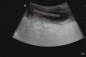
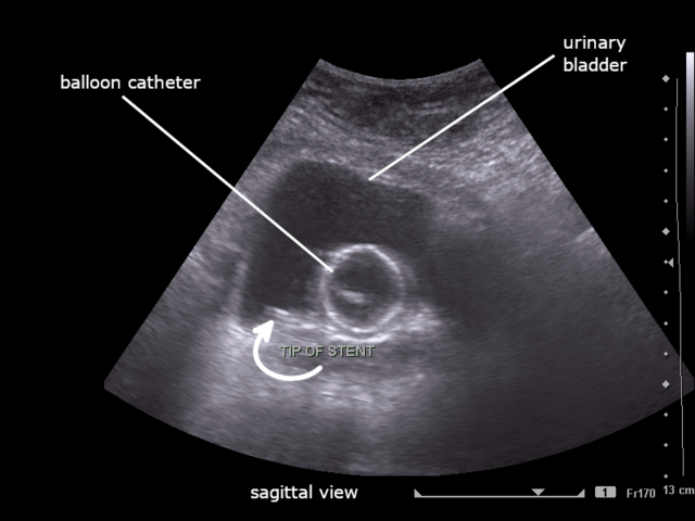
File:Ultrasound showing tip of ureteric stent within the urinary bladder.png  - Wikimedia Commons
File:Ultrasound showing tip of ureteric stent within the urinary bladder.png - Wikimedia Commons

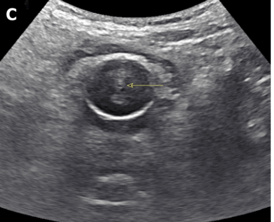
The 'ring of fire' Foley balloon – NephroPOCUS

Bladder ultrasound for catheterization and suprapubic aspiration (Chapter  16) - Pediatric Emergency Critical Care and Ultrasound
Bladder ultrasound for catheterization and suprapubic aspiration (Chapter 16) - Pediatric Emergency Critical Care and Ultrasound
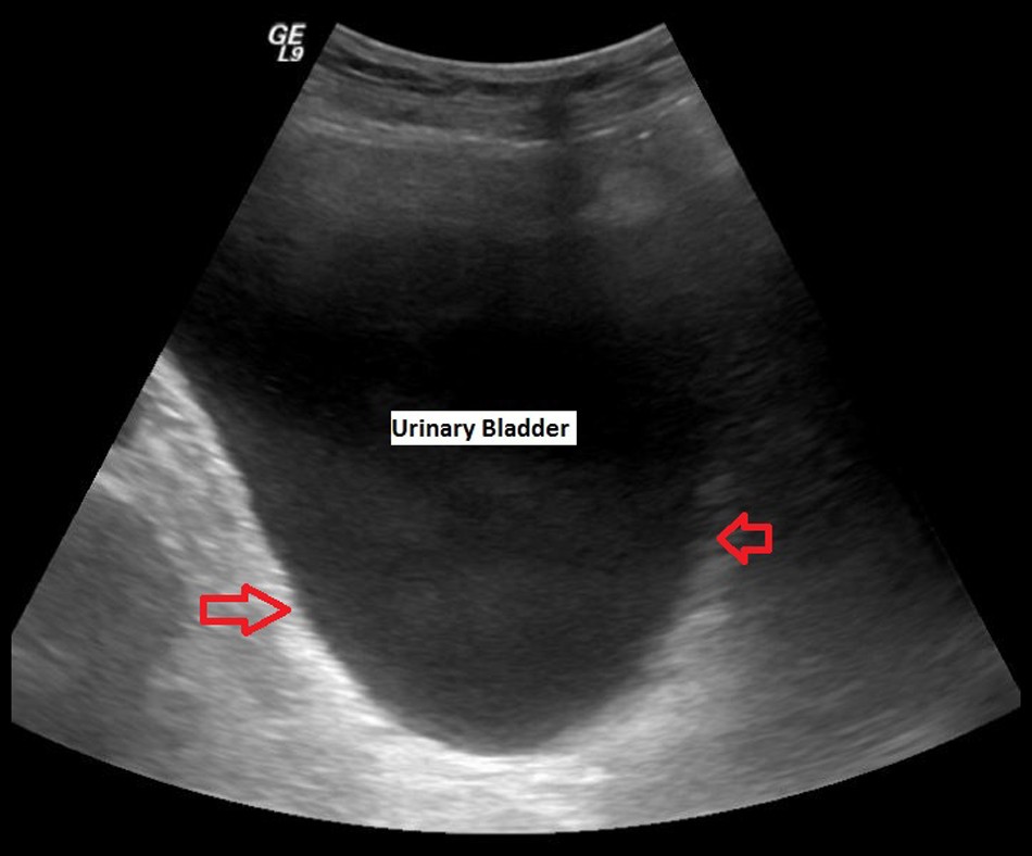
Bladder Ultrasound Made Easy: Step-By-Step Guide - POCUS 101

Holy Moly! What's Up with the Foley? - Renal Fellow Network

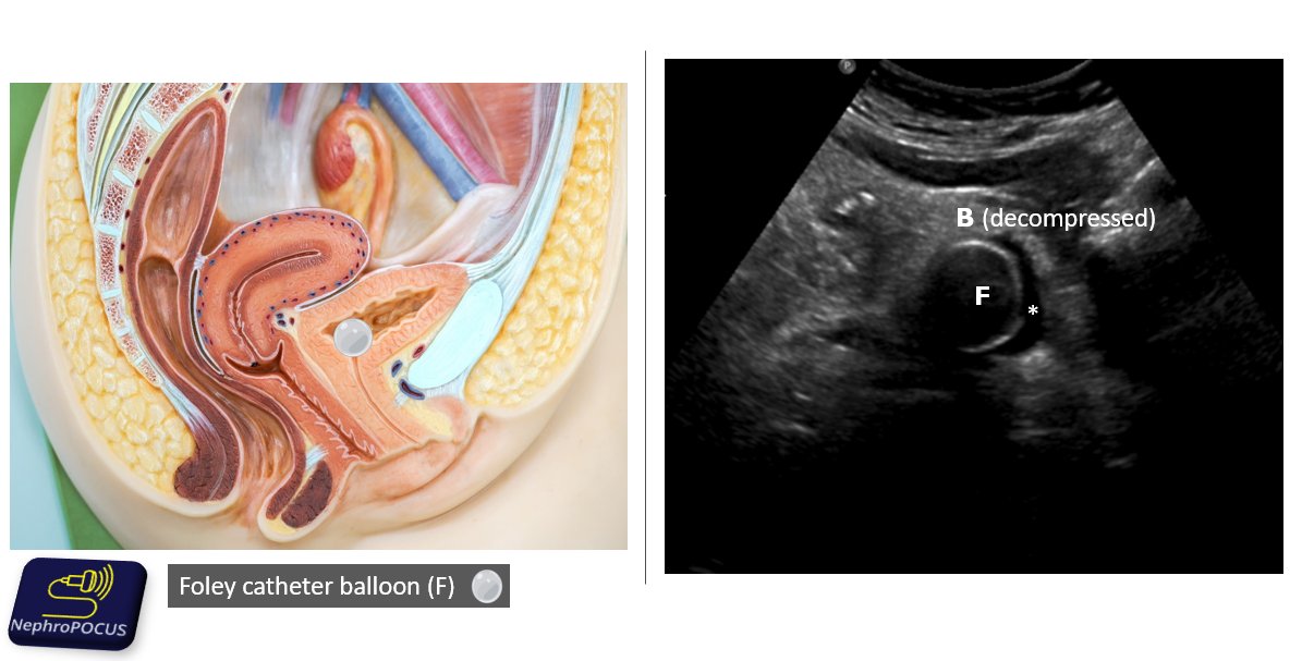
NephroPOCUS on X: "2. Urinary bladder decompressed by Foley catheter. As  the Foley balloon is filled with water, it appears as a well-defined cystic  structure on #POCUS Asterisk (*) indicates the tiny
NephroPOCUS on X: "2. Urinary bladder decompressed by Foley catheter. As the Foley balloon is filled with water, it appears as a well-defined cystic structure on #POCUS Asterisk (*) indicates the tiny
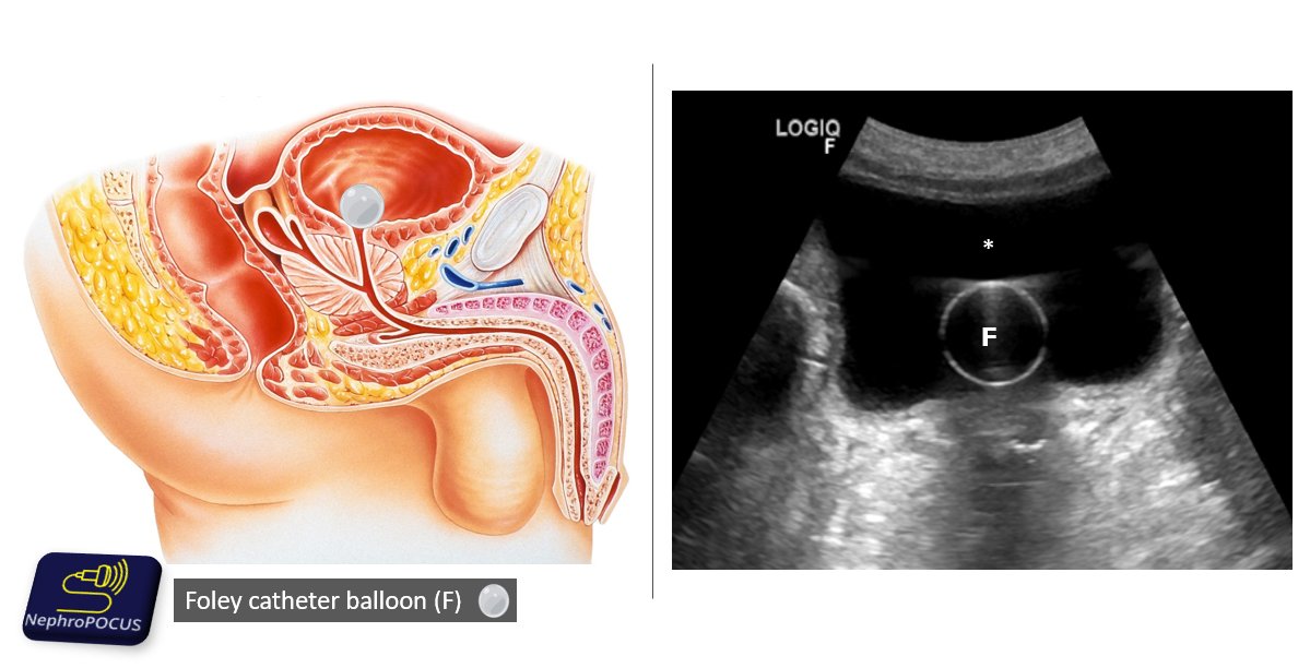
NephroPOCUS on X: "3. The sonographic image demonstrates the transverse  view of the bladder distended with urine(*) despite having a Foley in  place. Indicates obstruction of the catheter by a blood clot
NephroPOCUS on X: "3. The sonographic image demonstrates the transverse view of the bladder distended with urine(*) despite having a Foley in place. Indicates obstruction of the catheter by a blood clot

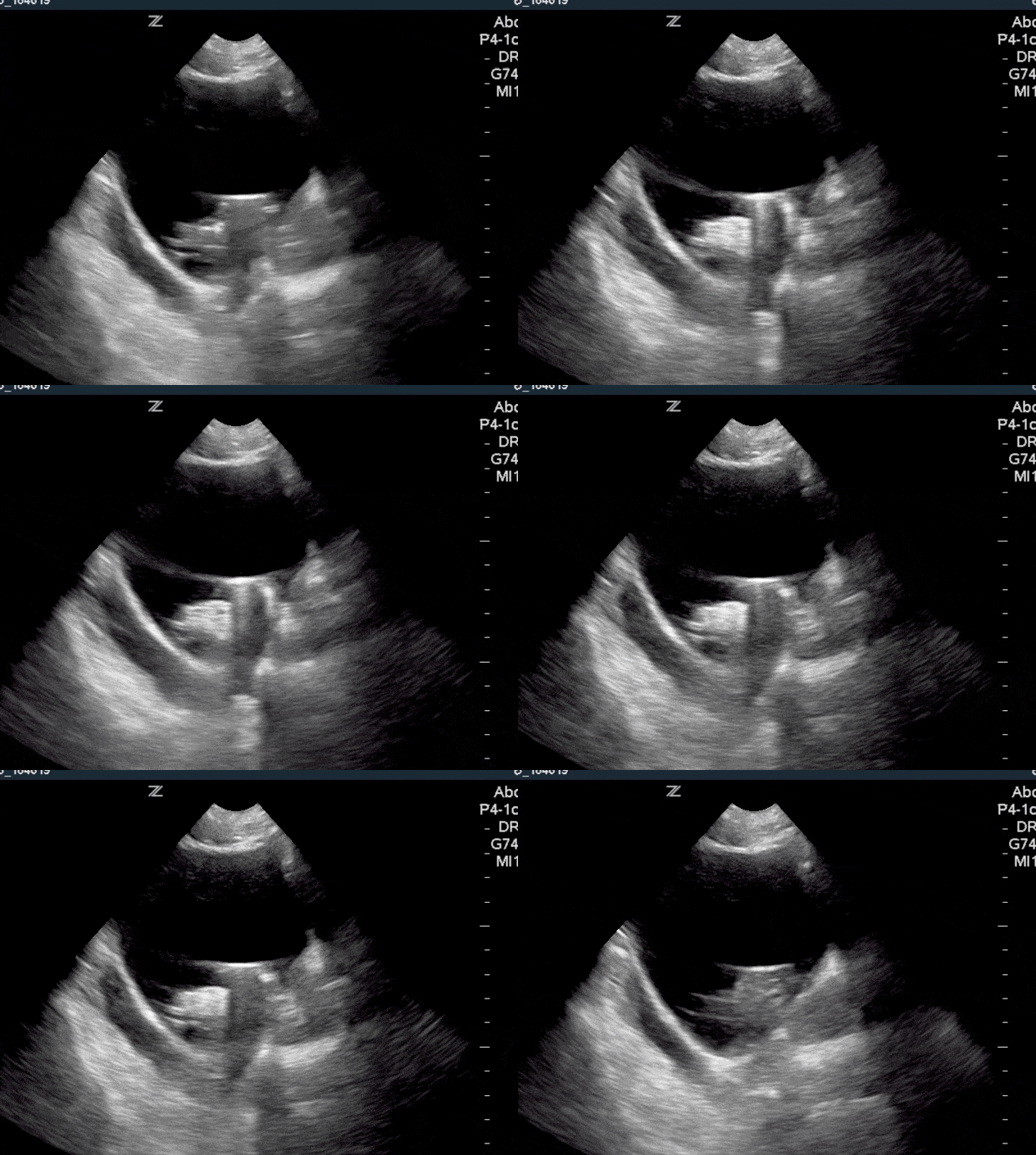
Sonography - Urinary Catheter - YouTube

Novel Use of Hydrodissection for the Insertion of Suprapubic Catheter Under  Ultrasound Guidance - ScienceDirect
Novel Use of Hydrodissection for the Insertion of Suprapubic Catheter Under Ultrasound Guidance - ScienceDirect

Obstructed Foley catheter – NephroPOCUS

Bladder Ultrasound Made Easy: Step-By-Step Guide - POCUS 101

Inadvertent placement of a urinary catheter into the ureter: A report of 3  cases and review of the literature. - Abstract - Europe PMC
Inadvertent placement of a urinary catheter into the ureter: A report of 3 cases and review of the literature. - Abstract - Europe PMC
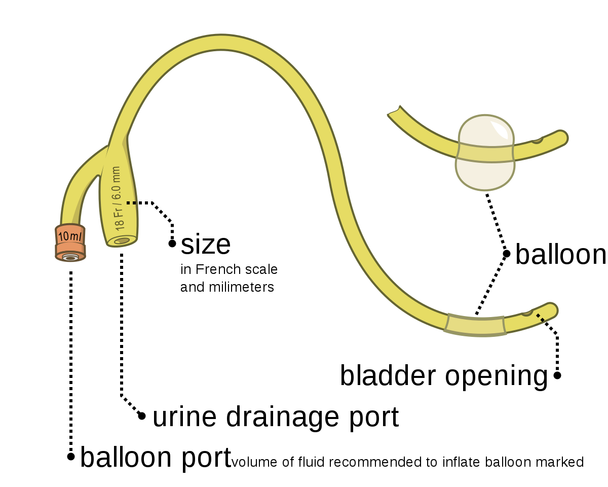
Foley catheter - Wikipedia

Persistent urine leakage around a suprapubic catheter: the experience of a  person with chronic tetraplegia | Spinal Cord Series and Cases
Persistent urine leakage around a suprapubic catheter: the experience of a person with chronic tetraplegia | Spinal Cord Series and Cases

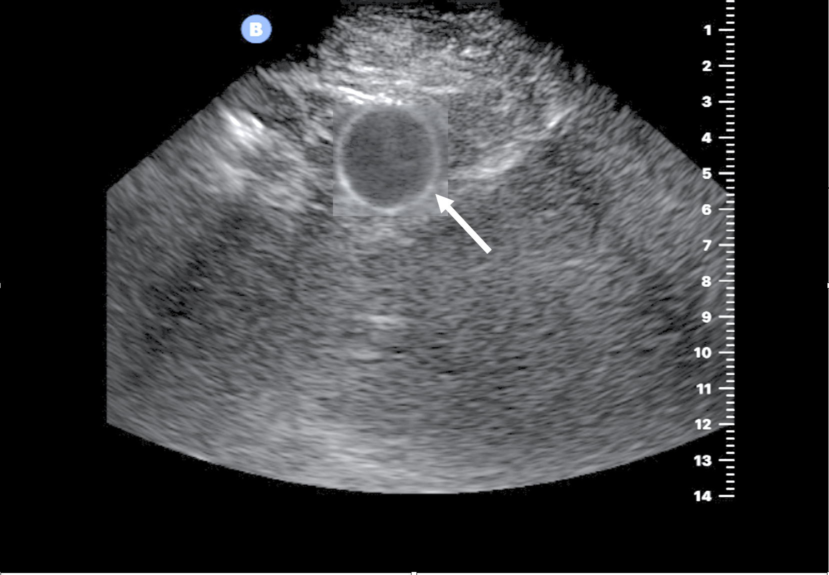
Misplaced Foley in the prostatic urethra – NephroPOCUS

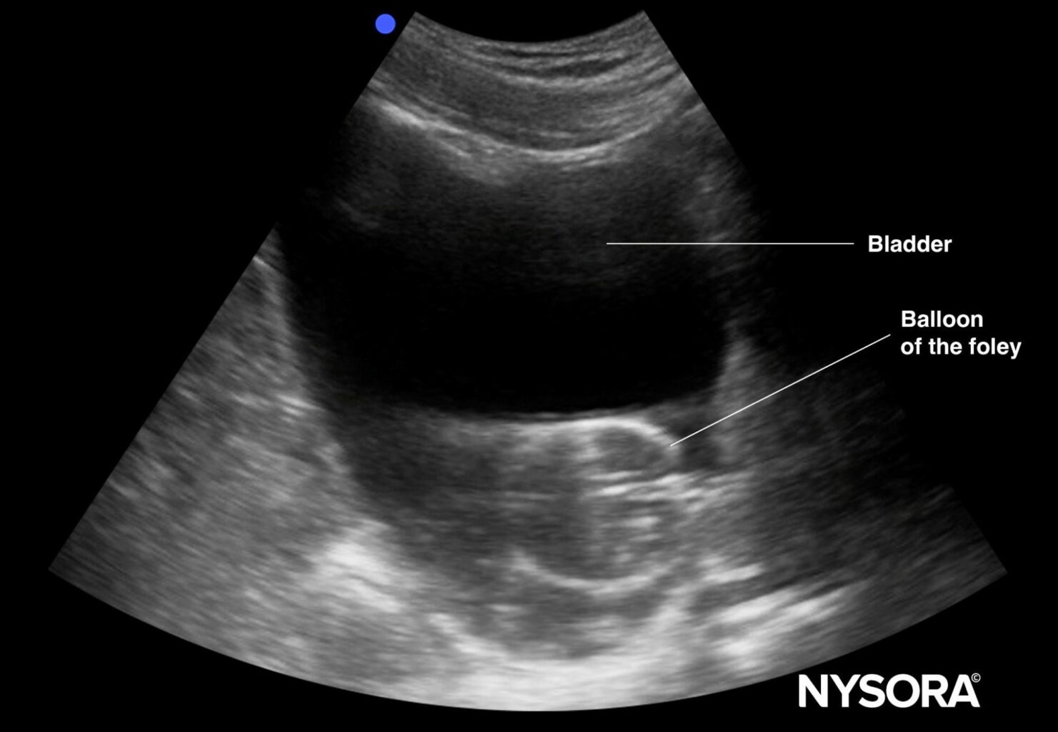
Ultrasound for Foley Catheter: Positioning & Obstruction Identification -  NYSORA
Ultrasound for Foley Catheter: Positioning & Obstruction Identification - NYSORA

POCUS Gallery - Renal Fellow Network

Ultrasound Guided Tips - ___Findings:- . *** Both Kidneys: _Marked  dilatation of both pelvicalyceal systems and ureters. _Renal sinus fat is  almost lost (in RED).. _Parenchymal thickness (in BLUE) measures about 10
Ultrasound Guided Tips - ___Findings:- . *** Both Kidneys: _Marked dilatation of both pelvicalyceal systems and ureters. _Renal sinus fat is almost lost (in RED).. _Parenchymal thickness (in BLUE) measures about 10

Obstructed Foley catheter – NephroPOCUS

Percutaneous puncture of urinary catheter balloon | Radiology Case |  Radiopaedia.org
Percutaneous puncture of urinary catheter balloon | Radiology Case | Radiopaedia.org

Bladder Ultrasound | Thoracic Key

Transabdominal ultrasound after emptying the bladder. The catheter... |  Download Scientific Diagram
Transabdominal ultrasound after emptying the bladder. The catheter... | Download Scientific Diagram